-ˋˏ ༻ 1 ༺ ˎˊ-
The Meeting of Elites
⟁
The elite meet to discuss the visitors and the results of the simulation
Centuries of observation has shown there are five docile personality types that exist in Organelle and five hostile ones
Some more intelligent than others
Docile personality types:
⟁
Totipotent:
Totipotent cells undergo zygotic genome activation (ZGA), which involves the synthesis of new transcripts
Totipotent cells undergo changes in DNA methylation, histone modifications, and chromatin remodeling
⟁
Pluripotent:
Pluripotent stem cells can differentiate into the ectoderm, mesoderm, and endoderm, which can give rise to all cell types
⟁
Multipotent:
unspecialized meaning they have not yet “decided” what type of adult cell they will be. They can self-renew and make two new stem cell. They can differentiate to make multiple types of cells
⟁
Oligopotent:
Oligopotent stem cells have a more restricted differentiation potential than multipotent or pluripotent stem cells
Oligopotent stem cells can differentiate into cells of the blood and immune systems. For example, myeloblast stem cells produce white blood cells, and lymphoid stem cells differentiate into lymphocyte
⟁
Unipotent:
Unipotent stem cells can only differentiate into one cell type
Unipotent stem cells are found in adult organ tissues that are dedicated to a specific cell lineage
⟁
Hostile Personalities:
Innate immunity
⟁
Adaptive immunity
⟁
Autoimmunity
⟁
Humoral immunity
⟁
Cell-mediated immunity
𖤓
━━━━━━━•°•°•❈•°•°•━━━━━━━
⟁
The Walk of Virtue
Is a message for the people
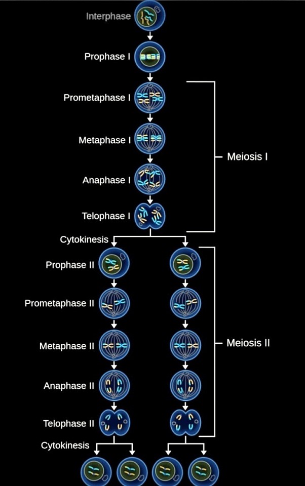
Meiosis
Telophase Two
Chromosomes gather: Chromosomes move to opposite poles of the cell, where they gather into two sets
Nuclear membranes form: A membrane forms around each set of chromosomes, creating two new nuclei
Chromosomes decondense: Chromosomes return to their "stringy" form
Cell division: The cell pinches in the middle and divides again, creating four daughter cells
Daughter cells are haploid: Each daughter cell has half the number of chromosomes as the original cell.
Daughter cells have new gene mixtures: Each daughter cell has a new combination of genes due to recombination during meiosis
𖤓
Anaphase Two
Anaphase II is a stage of meiosis II where sister chromatids separate and move to opposite poles of the cell
Centromere splitting: The centromere of each chromosome splits
Chromatid separation: Sister chromatids separate at the centromere
Microtubule attachment: Microtubules from the spindle attach to the kinetochore of each sister chromatid
Chromatid movement: Microtubules pull the sister chromatids to opposite poles of the cell
Cellular checkpoint: A checkpoint ensures that chromosomes formed after meiosis I have not changed
Cell elongation: Microtubules not attached to the kinetochore pull apart to elongate the cell
𖤓
Metaphase Two
Metaphase II is a stage of meiosis II where chromosomes line up in the middle of a cell to form a metaphase plate
Chromosomes line up: Chromosomes line up in pairs of sister chromatids along the metaphase plate
Microtubules attach: Microtubules from the centrosomes at opposite poles of the cell attach to the kinetochores of each chromosome
Centrioles at opposite poles: Centrioles are located at opposite poles of each daughter cell
Metaphase plate forms: The chromosomes align at the equator of the cell to form the metaphase plate
𖤓
Prometaphase Two
Prometaphase II is a stage of meiosis II when the nuclear envelope breaks down and the spindle is fully formed. During this stage, each sister chromatid forms a kinetochore that attaches to microtubules from opposite poles
Nuclear envelope breakdown: The nuclear membrane breaks down into small vesicles, allowing spindle microtubules to access the cell's genetic material
Kinetochore formation: A protein structure called a kinetochore forms around the centromere, which is the central point of the sister chromatids
Microtubule attachment: Microtubules from the centrosomes at the poles of the spindle attach to the kinetochores
Chromosome movement: The chromosomes move back and forth until they align on the metaphase plate in the center of the spindle
𖤓
Prophase Two
Prophase II is a stage of cell division in meiosis that involves the condensation of chromosomes and the breakdown of the nuclear envelope
Chromosomes condense: Chromosomes condense into visible X-shaped structures
Nuclear membrane dissolves: The nuclear membrane breaks down, allowing the chromosomes to become visible
Spindle fibers form: Centrioles migrate to opposite ends of the cell and begin to form spindle fibers.
Chromosomes move: The chromosomes begin to move toward the equator of the cell.
Kinetochore proteins assemble: Kinetochore proteins assemble on the outer chromatids of each chromosome
Meiotic spindle attaches: The meiotic spindle attaches to the kinetochores.
𖤓
Telophase One
Telophase I is a stage of meiosis where chromosomes gather at the poles of a cell, and the cell prepares to divide into two daughter cells
Homologous chromosomes separate: Homologous chromosomes separate and move to opposite sides of the cell
Nuclear envelope reforms: The nuclear envelope reforms around each set of chromosomes, creating two new nuclei
Cytokinesis occurs: The cell pinches in the middle, forming two daughter cells
Chromosomes decondense: The chromosomes de-condense back into chromatin
Spindle apparatus disappears: The spindle apparatus disappears
Daughter cells are not identical: The daughter cells are not identical because crossing over occurs, making each chromosome unique
𖤓
Anaphase One
Anaphase 1 is the third stage of meiosis I, and is characterized by the separation of homologous chromosomes
Homologous chromosome separation: Homologous chromosomes separate and move to opposite ends of the cell
Microtubule activity: Microtubules shorten and lengthen, pulling and pushing chromosomes and centrosomes
Cell elongation: The cell elongates as it prepares to divide
Spindle apparatus: The spindle apparatus moves chromosomes to the poles of the cell
Kinetochore microtubules: Kinetochore microtubules shorten, pulling homologous chromosomes to opposite poles
Non-kinetochore microtubules: Non-kinetochore microtubules lengthen, pushing centrosomes apart
𖤓
